
Ruptured eardrum Symptoms, causes, and treatments
Browse 1,300+ eardrum stock photos and images available, or search for perforated eardrum to find more great stock photos and pictures. perforated eardrum Sort by: Most popular Ruptured (perforated) eardrum Ruptured eardrum. Anatomy of the humans eardrum. Healthy and perforated tympanic membrane.
Ruptured Eardrum Doctor Philadelphia
5 Additional images. 6 References. 7 External links. Toggle the table of contents.. In the anatomy of humans and various other tetrapods, the eardrum, also called the tympanic membrane or myringa, is a thin, cone-shaped membrane that separates the external ear from the middle ear.

Researchers Create Scaffolds For Eardrum Replacement Minds of Malady
Browse 1,300+ ear drum stock photos and images available, or search for inner ear or ear canal to find more great stock photos and pictures. inner ear ear canal cochlea otitis media doctor throat nostril middle ear hearing ear anatomy Sort by: Most popular Ruptured (perforated) eardrum Ruptured eardrum. Anatomy of the humans eardrum.
Ear Infection (Middle Ear) Causes, Symptoms, Diagnosis and Treatment
Awesome prices & high quality here on Temu. New users enjoy free shipping & free return. Don't swipe away. Massive discounts on our products here - up to 90% off!
Ear Discharge Is It dangerous? Living Herself
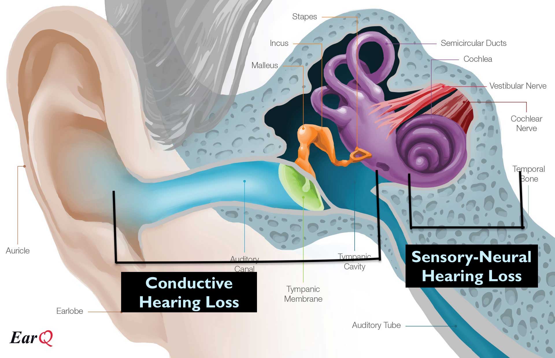
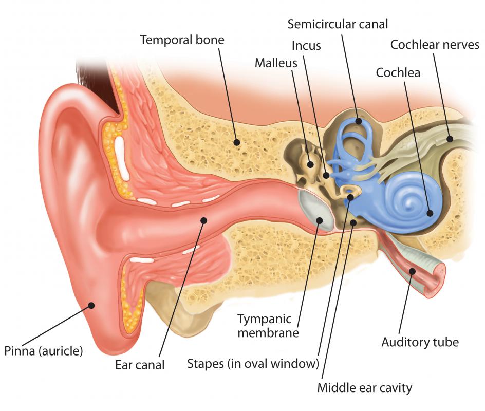
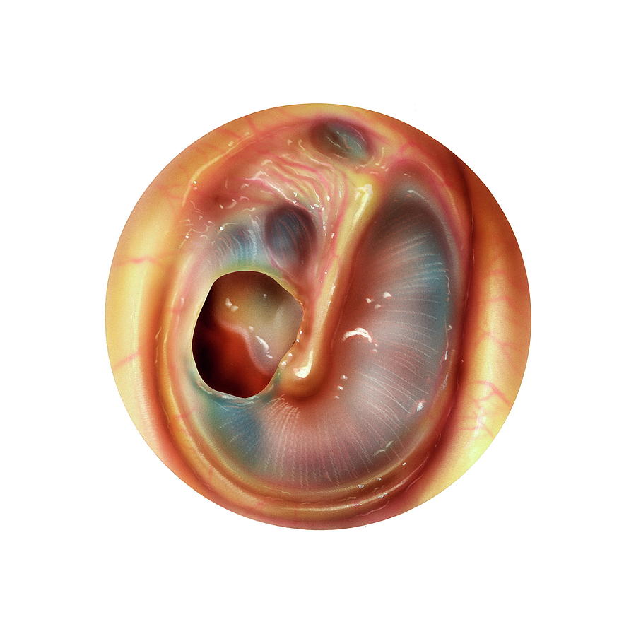
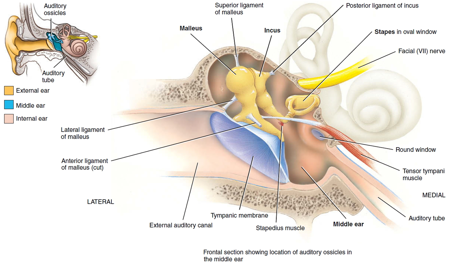
The eardrum (tympanic membrane) is the circular surface that dominates the image. The eardrum vibrates from sound waves. The vibration of the ear drum has to be transferred to the inner ear to produce electrical signals that we interpret as sound. The transfer of the vibrating eardrum to the inner ear is accomplished by three bones in the.

Myringoplasty (repairing a hole in the ear drum) Dr Bridget Clancy
The image is of a normal nasopharynx and the opening to the Eustachian tube. The Eustachian tube goes from the back of the nose (nasopharynx) to the middle ear. Normally the tube remains closed and opens when you swallow, yell or pop your ear with a Valsalva Maneuver. See appendix I: How to "pop" your ears. Ear Anatomy
Eardrum Ear canal Outer ear Middle ear, ear, hand, people, human png
OTOVEL® (ciprofloxacin and fluocinolone acetonide) is used in children 6 months of age and older, who have a tiny cylinder tube in their eardrum known as a tympanostomy tube to prevent excess fluid in the middle ear. Otovel is used to treat a type of middle ear infection called acute otitis media with tympanostomy tubes (AOMT) caused by.
What is a Ruptured Eardrum? (with pictures)
Pictures of Different Ear Abnormalities by Dr. Christopher Chang, last modified on 6/17/21. One of the most common reasons for a patient to see an ENT doctor are issues related to the ear, especially because the ear is not something that can be easily visualized at home.
/GettyImages-eardrum-047cd39dca7f40afaefb3a973c268925.jpg)
Eardrum Anatomy, Function, and Treatment
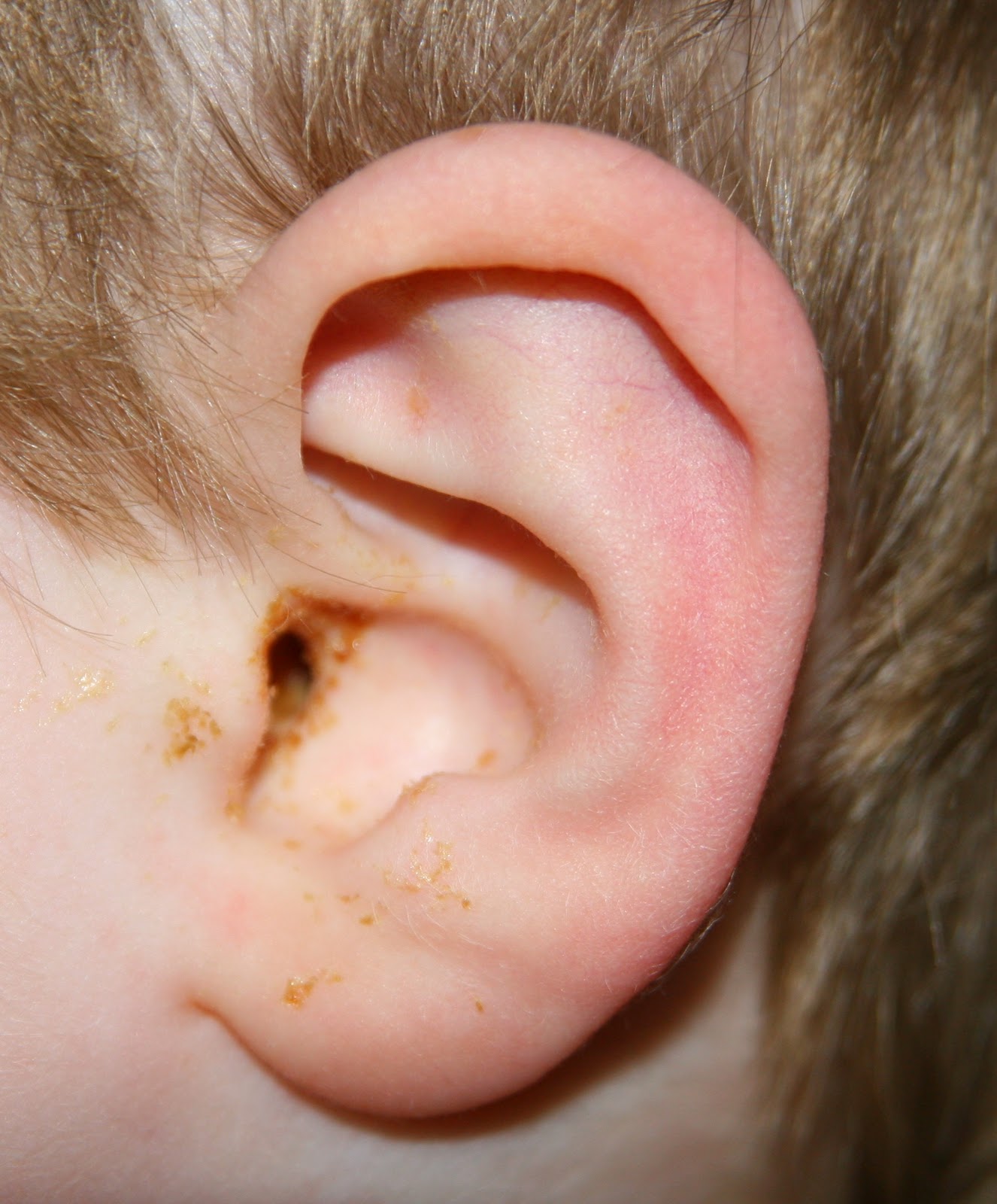
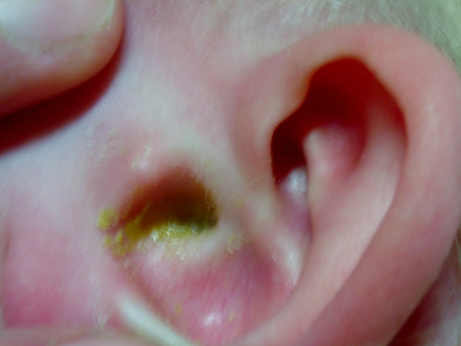
Middle Ear Infection Images Six year old with an early ear infection. He had complained of ear pain for three to four hours. Red dilated blood vessels at the upper part of the ear drum. Seventeen year old male with a two day history of ear pain and sore throat.

Eardrum Wikipedia
Fluid in the ear Images. Five year old female with fluid in the middle ear. The air bubbles are easily seen and the fluid is thin and watery looking. Most likely the fluid will clear over the next few days. Seven year old male, with a note from teacher and school nurse, stating that his grades have fallen off over the past two to three months.
Perforated Eardrum Photograph by Bo Veisland/science Photo Library
Inner ear: The inner ear, also called the labyrinth, operates the body's sense of balance and contains the hearing organ. A bony casing houses a complex system of membranous cells. The inner ear.
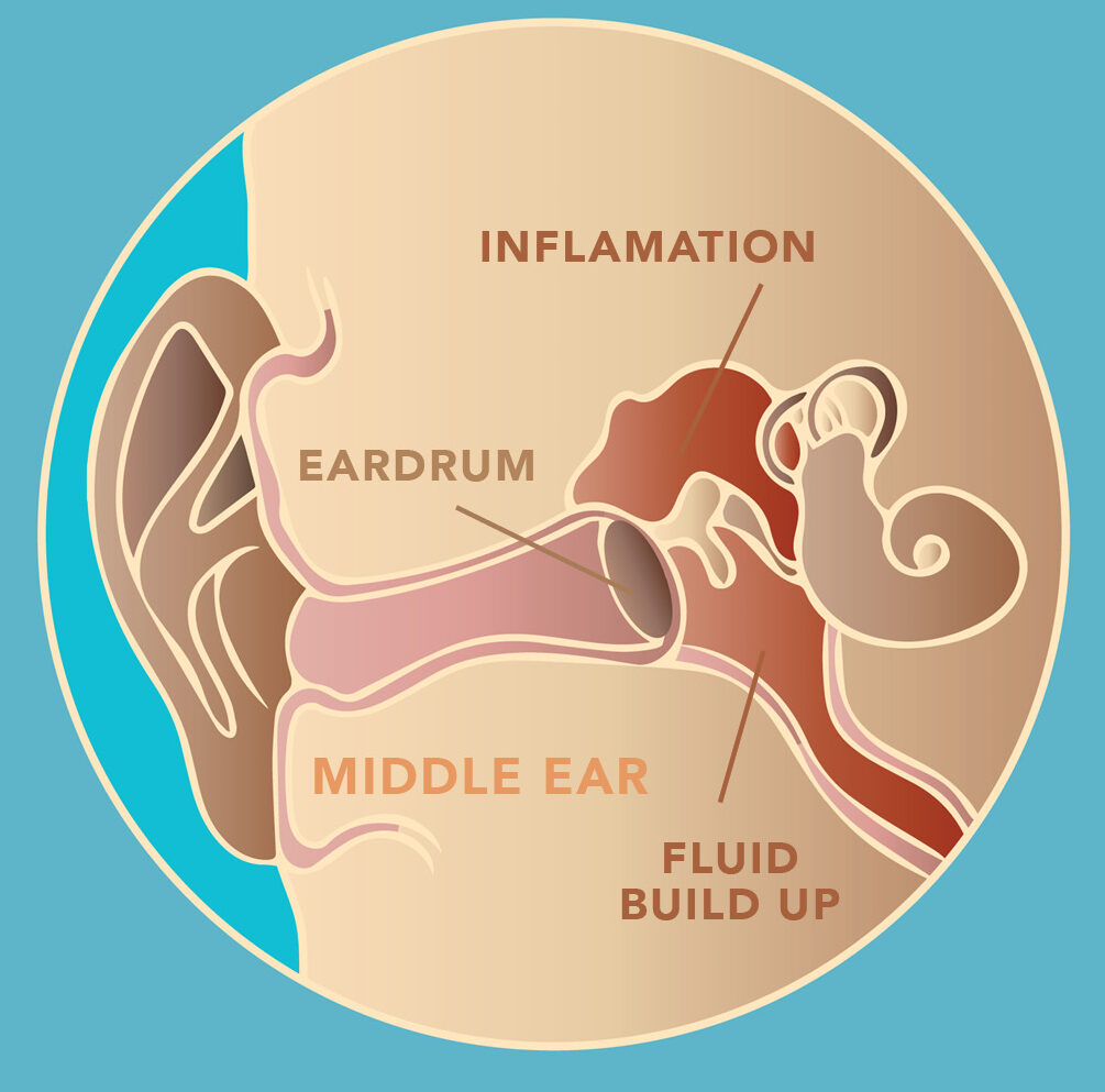
What a Middle Ear Infection Looks Like PhotoniCare
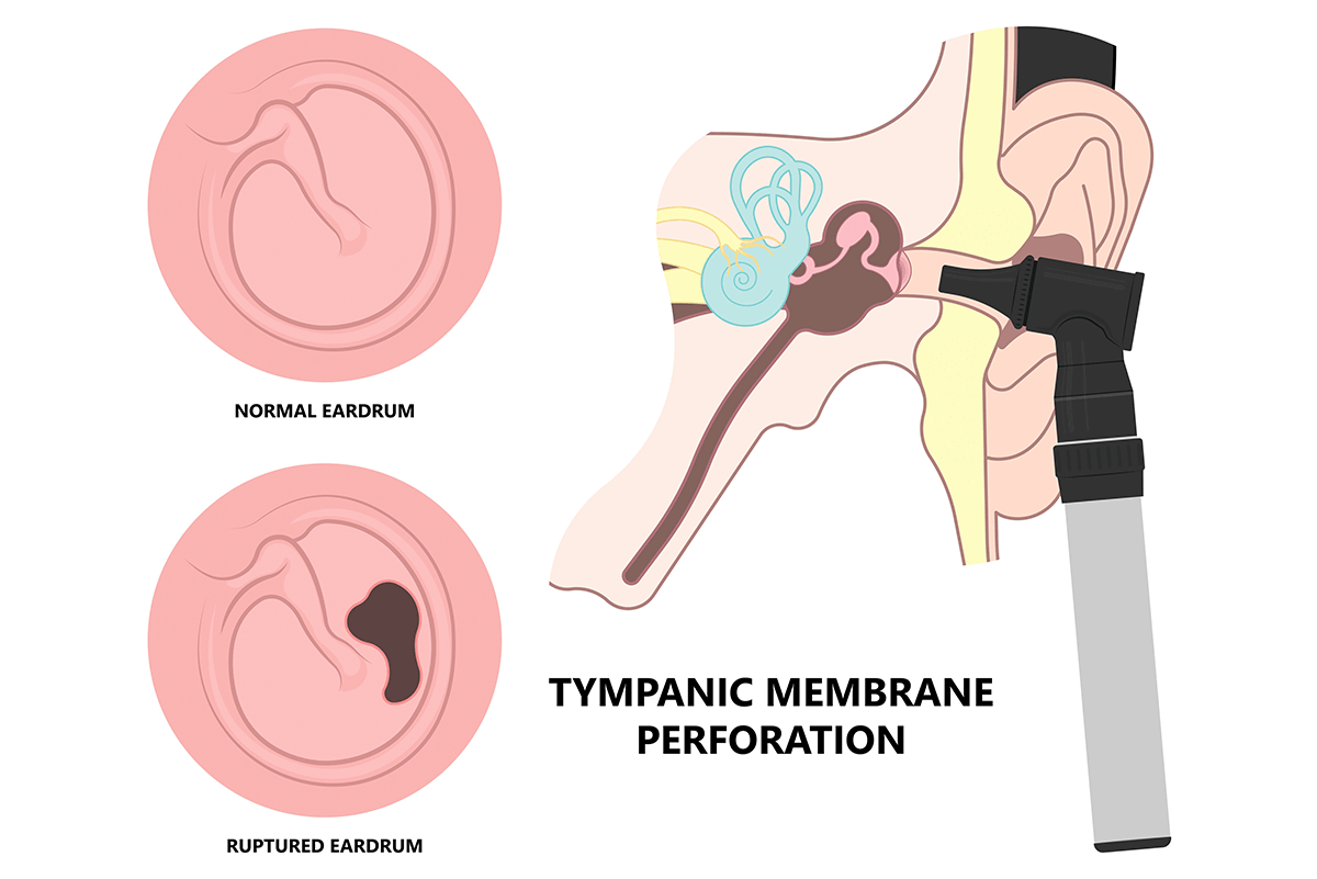
Tympanic Membrane (Eardrum) Your tympanic membrane (eardrum) is a thin, circular layer of tissue that separates your outer ear from your middle ear. Your eardrum plays an important role in hearing. It also protects your middle ear from dirt, bacteria and debris. Contents Overview Function Anatomy Conditions and Disorders Care Additional Common.

Ruptured eardrum causes, signs, symptoms, diagnosis & treatment
Browse 526 eardrum photos and images available, or search for perforated eardrum to find more great photos and pictures. 9 Browse Getty Images' premium collection of high-quality, authentic Eardrum stock photos, royalty-free images, and pictures. Eardrum stock photos are available in a variety of sizes and formats to fit your needs.
Ear Drum Anatomy, Causes, Diagnosis & Treatment for Busted Ear Drum
Anatomy The eardrum has three layers: the outer layer, inner layer, and middle layer. The middle layer is made of fibers that give the eardrum elasticity and stiffness. Cartilage holds the eardrum in place. The eardrum covers the end of the external ear canal and looks like a flattened cone with its tip pointed inward toward the middle ear.
Ruptured eardrum (perforated eardrum) Disease Reference Guide
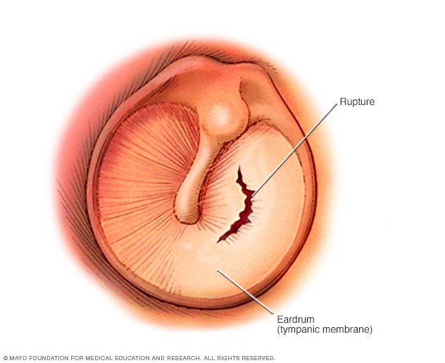
Overview A ruptured eardrum (tympanic membrane perforation) is a hole or tear in the thin tissue that separates the ear canal from the middle ear (eardrum). A ruptured eardrum can result in hearing loss. It can also make the middle ear vulnerable to infections. A ruptured eardrum usually heals within a few weeks without treatment.
Everything Kristi! Burst Ear Drum
The ear canal, or auditory canal, is a tube that runs from the outer ear to the eardrum. The ear has outer, middle, and inner portions. The ear canal and outer cartilage of the ear make up the.